Introduction
Filler injections are often promoted as a quick and simple way to enhance the nose without surgery. While most patients experience no serious problems, vascular complications can occur when filler blocks the blood supply to the skin. Although rare, this is one of the most challenging situations in rhinoplasty because damaged skin cannot simply be replaced with another implant.
This case demonstrates how complex nasal reconstruction can restore both appearance and structural support after filler-induced skin necrosis.
Understanding Filler Necrosis
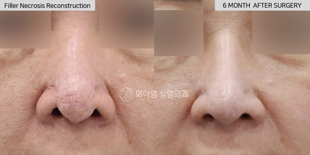
This 43-year-old woman received a filler injection into the nasal tip approximately five years earlier. Her goal was simple: to create a slimmer, slightly more projected nasal tip without surgery.
Unfortunately, the filler compromised the blood supply to the nasal tip, resulting in skin necrosis. Although the acute wound eventually healed, the damage left behind permanent scar contracture, loss of healthy soft tissue, and significant skin deficiency over the tip of the nose.
Patients with this type of injury often notice that the nasal tip becomes firm, tight, and distorted over time. In many cases, the skin no longer has enough flexibility to support conventional revision rhinoplasty techniques.
Why Reconstruction Is Different
Revision rhinoplasty after filler necrosis is fundamentally different from routine cosmetic revision surgery.
The challenge is not simply changing the shape of the nose. The surgeon must rebuild a nose that has lost healthy tissue.
Years after necrosis, dense scar tissue often replaces the normal soft tissue. Blood circulation may also be less reliable than in an untouched nose. In addition, the skin covering the nasal tip is frequently thinner, tighter, and less forgiving because part of the original tissue has already been lost.
For these reasons, reconstruction must prioritize long-term tissue survival and structural stability before cosmetic refinement.
How We Reconstructed the Nose
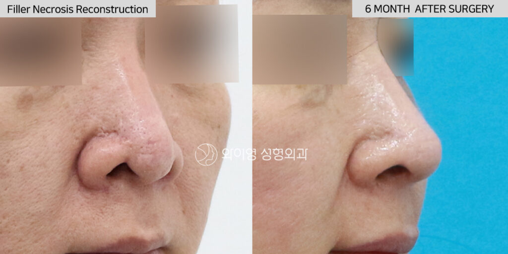
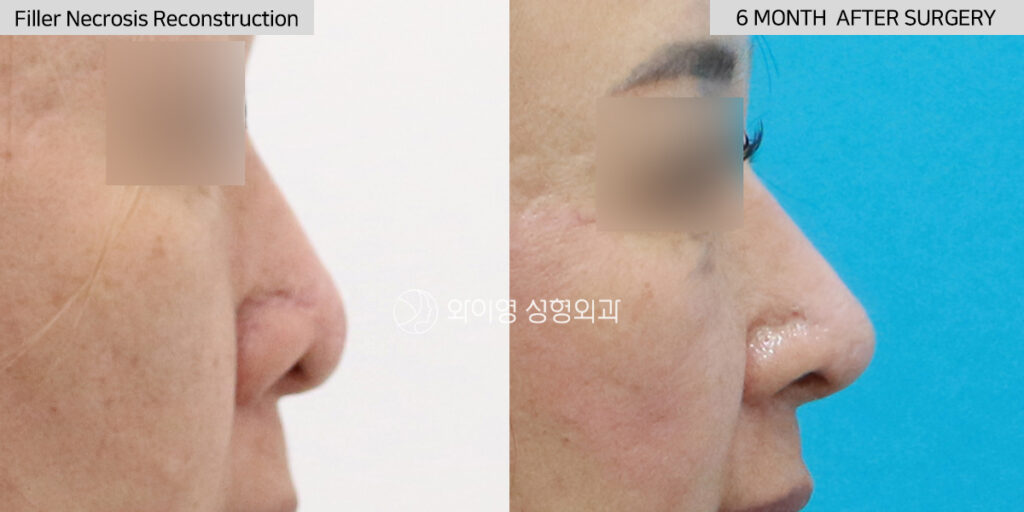
For this patient, we chose autologous rib cartilage to reconstruct both the nasal bridge and the nasal tip.
Because the soft tissue had already been compromised by previous necrosis, we wanted to avoid introducing another permanent synthetic implant. Using the patient’s own rib cartilage provided a biologically compatible framework with excellent strength and a lower long-term risk of complications.
The remaining skin deficiency at the nasal tip was reconstructed using a composite ear graft, which supplied both cartilage and skin to restore the damaged soft tissue.
This combination allowed us to rebuild the structural foundation of the nose while creating sufficient support for a natural nasal contour.
Most importantly, the reconstruction was designed with tissue safety as the highest priority rather than simply maximizing projection.
Why We Chose Rib Cartilage
Autologous rib cartilage is often considered the most reliable graft material for complex revision rhinoplasty when significant structural reconstruction is required.
Unlike silicone implants or other synthetic materials, rib cartilage becomes part of the patient’s own tissue. It provides enough strength to support the reconstructed nose while minimizing the risk of future implant-related complications.
In cases involving previous skin necrosis, scar tissue, or tissue deficiency, rib cartilage offers several important advantages:
- Excellent structural strength for rebuilding the nose
- No permanent synthetic implant beneath compromised skin
- Better long-term stability in difficult revision cases
- Sufficient volume for complete reconstruction of both the bridge and the nasal tip
While not every revision rhinoplasty requires rib cartilage, it is frequently the safest and most predictable option when healthy tissue has already been lost.
Frequently Asked Questions
Can filler really cause skin necrosis?
Yes. Although uncommon, filler can block the blood vessels supplying the nasal skin. Without adequate blood flow, skin tissue may become damaged or die, requiring reconstructive surgery.
Is revision surgery possible years after filler necrosis?
Yes. Reconstruction can often be performed even years later. However, scar tissue and skin quality must be carefully evaluated before surgery.
Why not use another silicone implant?
When the skin has already been weakened by previous necrosis, placing another permanent implant may increase the risk of future complications. In these situations, autologous rib cartilage is often a safer reconstructive option.
Will the reconstructed nose look natural?
The primary goal is to restore healthy tissue and structural stability. Once a stable foundation has been created, a natural appearance usually follows.
How long does recovery take?
Most swelling gradually improves over several months, while the final result typically continues to mature over approximately one year.
Learn More About Revision Rhinoplasty
Filler-induced skin necrosis is one of the most challenging conditions in nasal reconstruction. Successful treatment requires more than cosmetic correction—it demands careful structural planning, healthy tissue management, and an individualized surgical approach.
Learn more about our philosophy and approach to complex revision rhinoplasty.
Conclusion
Filler necrosis is one of the most complex complications encountered in nasal reconstruction, but it does not necessarily mean that the nose cannot be restored.
Careful planning, healthy tissue management, and appropriate graft selection are essential for achieving both functional stability and a natural appearance.
In this patient, reconstruction using autologous rib cartilage and a composite ear graft allowed us to rebuild a nose that had suffered significant tissue loss, while prioritizing long-term safety and reliable structural support.
Related Articles
- Silicone Implant Extrusion Reconstruction in Korea
- Rib Cartilage Rhinoplasty Without Implants
- Silicone Implant vs Rib Cartilage for Dorsal Augmentation

